Detecting single nucleotide variations (SNVs) offers powerful actionable insights in diagnosing diseases, monitoring cancer progression and identifying pathogen variants. However, reliably spotting these minor changes in rare cell populations is technically challenging due to the required sensitivity and specificity. As a result, the lack of affordable, simple and highly specific techniques is limiting the ability to take full advantage of the opportunities in personalised medicine and advanced diagnostics.
Ongoing research is uncovering several promising strategies for detecting gene mutations that rely on ligases, such as Ampligase™.1 This thermostable DNA ligase joins two adjacent DNA strands that are hybridised to a complementary target, but only if they perfectly match at the joining site.
Ampligase is derived from a thermophilic bacterium, which means it is stable and active at much higher temperatures than conventional DNA ligases. It has a half-life of 48 hours at 65 °C and greater than an hour at 95 °C, and it is active for at least 500 thermal cycles (94 °C/80 °C) or 16 hours of cycling.2 This exceptional thermostability permits extremely high hybridisation stringency and ligation specificity.
Advantages of ligase-based techniques
Ligase-based techniques have several advantages over PCR, which has been the gold standard for DNA amplification. PCR is limited by the specificity of DNA polymerase in recognising single-base mismatches.1 By contrast, Ampligase’s specificity for exact matches with the template sequence allows SNVs to be detected with extreme accuracy. This makes it particularly effective in areas like cancer diagnostics and monitoring, where rare mutations must be detected among a background of wild-type DNA. 
In addition, PCR relies on temperature cycling, while Ampligase-based methods can operate under isothermal conditions, eliminating the need for sophisticated thermal cycling equipment.
Ampligase’s ability to join DNA strands with high specificity forms the backbone of several innovative methodologies designed to detect genetic biomarkers with incredible precision. This blog post delves into recent advances in this area, demonstrating the potential of Ampligase-based technologies in genetic analysis and disease diagnosis.
Enhancing CRISPR/Cas12a sensitivity with rolling circle amplification
Identifying genetic biomarkers is crucial in understanding the causes of diseases and developing therapies that can target them. Traditional methods like PCR can fall short in specificity, particularly in discriminating single-nucleotide variations critical for precision medicine.
In the quest for ultra-sensitive detection methods, a study by Cao et al. introduced an ingenious combination of CRISPR/Cas12a with rolling circle amplification (RCA).3 This method combines the high specificity of CRISPR/Cas12a to identify the mutation of interest, coupled with the amplification capabilities of RCA to enhance the detection sensitivity.
Rolling circle amplification (RCA) is a popular isothermal reaction because of its high efficiency, low error rate and simple operation. In this reaction, Ampligase is used to form a single-stranded DNA circle. The padlock probe is designed such that the 3' and 5' ends pair with separate halves of the target sequence. When the target gene and the probe are perfectly matched, Ampligase connects the 3'-terminal hydroxyl group and the 5'-terminal phosphate group of the probe to form a circular closed loop. 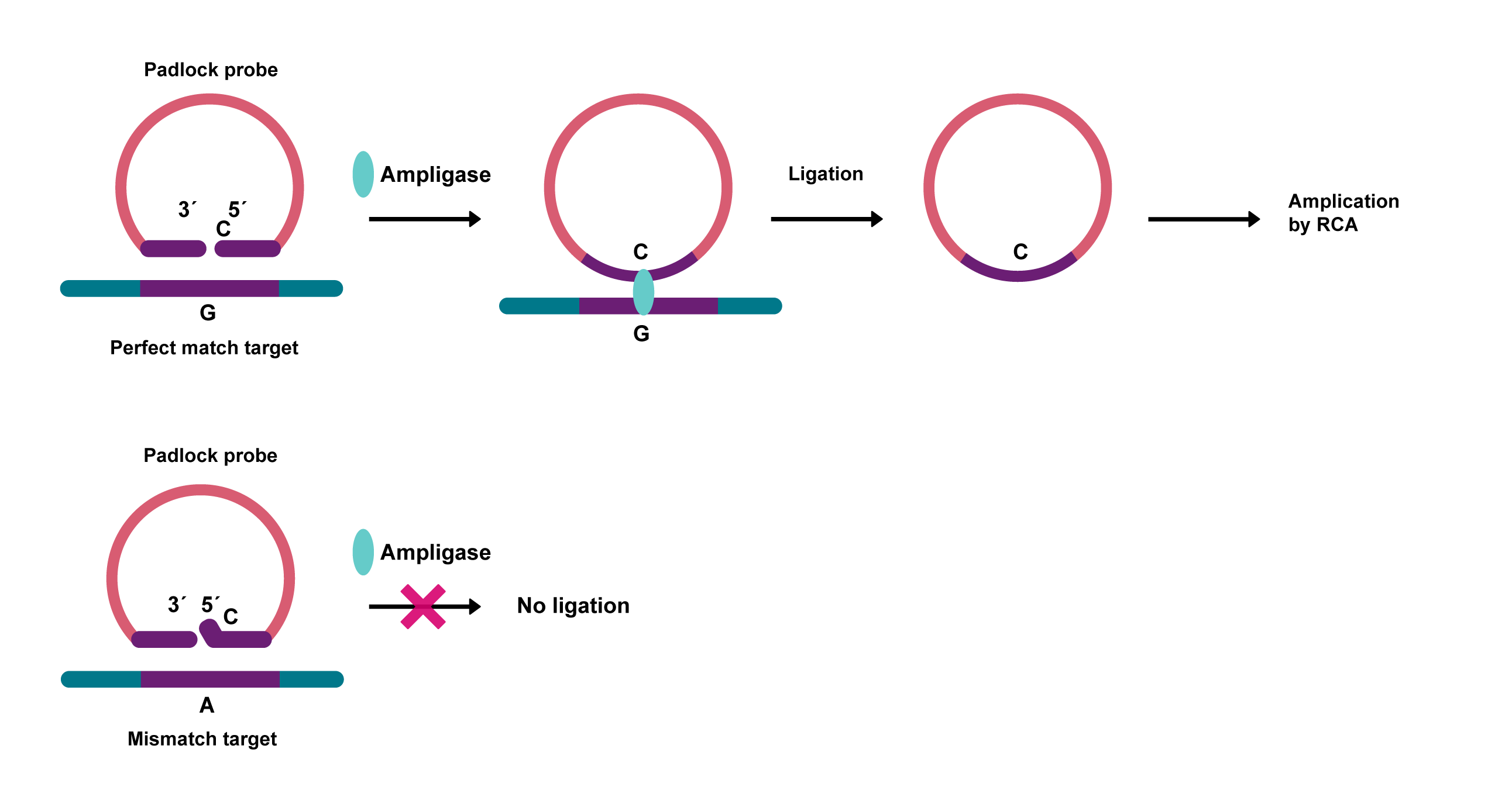
Figure 1. Padlock probe circularisation using Ampligase.
This critical ligation step ensures that only the specific mutation triggers amplification. RCA typically uses phi29 polymerase, as its high processivity and strand-displacement activity can amplify circular DNA templates, forming long, single-stranded DNA products.
The amplified product is then detected through the CRISPR/Cas12a system, offering a low detection limit and high specificity. On recognising the target DNA, Cas12a engages in non-specific trans cleavage, cutting all single-stranded DNA in the vicinity. By including reporter probes in the sample, the researchers could measure the signal as Cas12a freed the fluorescent dyes.
The researchers demonstrated this approach by detecting the PIK3CA H1047R mutation, which is closely linked to breast cancer. Given the mutation’s significance, its detection is crucial for early diagnosis, monitoring and treatment planning.
The study showed that the RCA-CRISPR/Cas12a method could detect PIK3CA H1047R at concentrations as low as 0.01 fM, accounting for only 0.036% of the mixture with wild-type samples. This highlights its potential as a straightforward, rapid and highly specific strategy for medical diagnostics.
Ultra-sensitive monitoring of leukaemia
In many cancers, such as acute myeloid leukaemia (AML), detecting minute quantities of mutant DNA can give earlier indications of how well a therapy is working. This precision can help doctors make better decisions about treatment and potentially improve patient survival. However, it is challenging to accurately identify rare mutations within a vast excess of normal DNA. Traditional methods, while useful, often fall short in sensitivity or specificity, particularly when mutations occur at extremely low frequencies.
Assessing the measurable residual disease following AML treatment can be done using PCR methods on bone marrow samples. However, as more targeted therapies become available, clinicians will need highly sensitive, specific and practical tests to fulfil the promise of a revolution in personalised medicine.
To enhance the precision of cancer monitoring and treatment, Chen et al. introduced superRCA assays, a novel technology that significantly improves the detection of rare tumour-specific mutations in patient samples.4
SuperRCA uses PCR to amplify DNA sequences known to be mutated in a patient’s cancerous cells. These amplified strands are circle ligated by Ampligase, which then go through RCA using phi29 polymerase. The RCA products are then analysed with padlock probes specific to the wild type or mutant sequences, which Ampligase ligates when there is a perfect match. These ligated padlock probes then provide templates for secondary RCA reactions, which can be detected using fluorescence-labelled hybridisation probes.
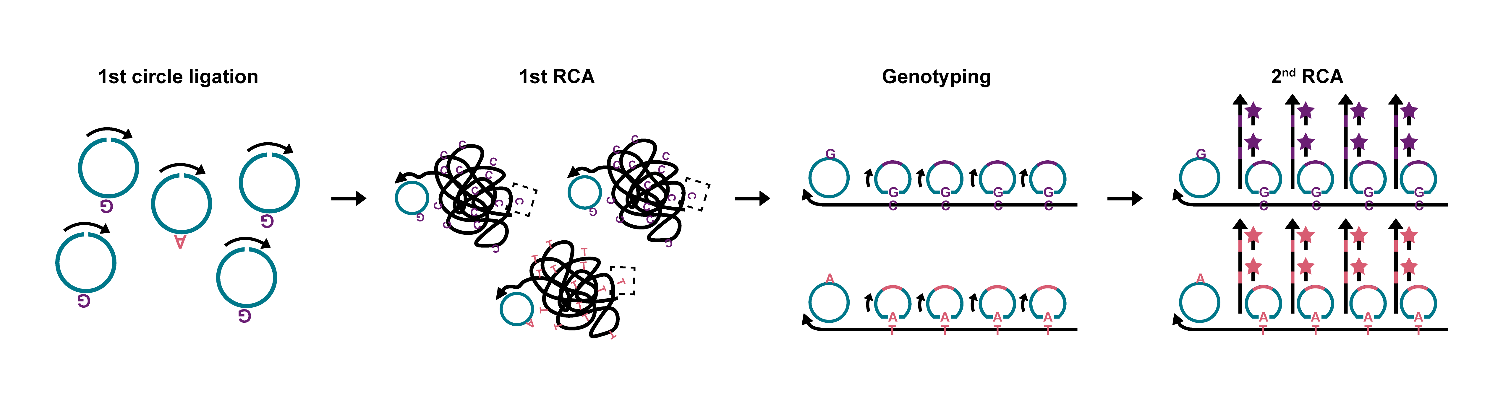
Figure 2. Schematic representation of the superRCA method, amplifying the sequences of interest for detection by flow cytometry. Adapted from Chen et al. under a Creative Commons licence.
This method generates highly specific, amplified products from minimal quantities of mutant DNA, enabling the detection of single-nucleotide mutations against a background of up to 100,000-fold more normal DNA. The sensitivity and specificity allow for monitoring disease progression and early detection of recurrence, offering a basis for timely therapeutic interventions. By comparison, the NGS limit of detection for SNVs is approximately 2-5%, limiting its use in identifying residual cancer.5,6
In one patient included in the study, NGS failed to detect a remaining cancerous mutation after initial treatment, leading to relapse. Later analysis with superRCA detected the lurking cells’ DNA, which could have altered the patient’s treatment pathway. While NGS can be adapted to be more sensitive than this approach, it comes at a much greater cost and turn-around time.
In addition, by detecting these mutations in blood samples rather than bone marrow, superRCA assays could significantly improve the patient experience and reduce the clinical burden of testing.
Detecting SARS-CoV-2 variants and HPV
The COVID-19 pandemic emphasised the importance of precise, sensitive and simple viral detection methods. In 2021, Wang et al. published an approach offering a rapid way to distinguish between wild-type and single-base mutations in the SARS-CoV-2 virus.7
Wang et al. built on the Ligase Chain Reaction (LCR), which exploits the DNA ligase enzyme to amplify DNA sequences with high fidelity. It requires minimal temperature cycling, simplifying the equipment needed. The researchers developed this further by using Pyrococcus furiosus Argonaute (PfAgo), a thermophilic DNA-guided enzyme that cleaves DNA longer than 14 bases.8 PfAgo can use the LCR products as DNA guides, cleaving a molecular beacon target that releases a fluorophore for detection.
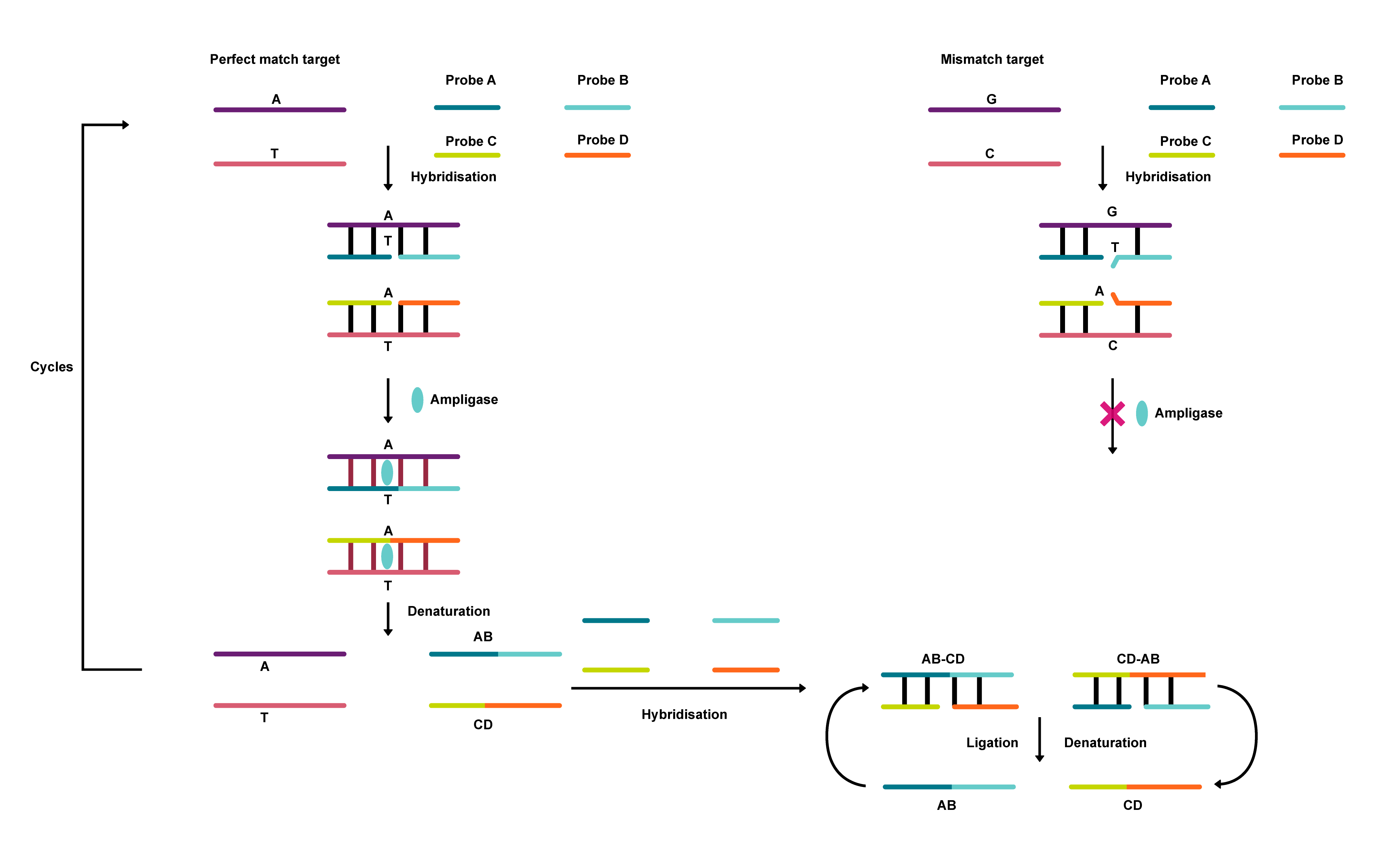 Figure 3. Flow diagram representing the process for the ligase chain reaction.
The technique, named PLCR (PfAgo coupled with modified Ligase Chain Reaction for nucleic acid detection), achieves 10 aM sensitivity for DNA or RNA detection in 70 minutes and can differentiate single base mutations effectively. The study showed that PLCR could be multiplexed to detect variants of both RNA and DNA viruses, demonstrated using SARS-CoV-2 and HPV. Detecting RNA required adding reverse transcriptase to the reaction.
PLCR combines PfAgo’s high-temperature DNA cleavage capacity with LCR’s single-base mutation discrimination ability, circumventing the complexities and limitations of prior approaches. Ampligase’s thermostability enables the reaction to process efficiently at elevated temperatures, which are optimal for PfAgo’s activity. Wang et al. found that Ampligase was better than Taq DNA ligase in terms of template-dependent amplification.
PLCR’s high sensitivity and specificity for detecting viral variants makes it a promising advance in molecular diagnostics, potentially enabling more timely and effective responses to outbreaks.
Electrochemical biosensor for point mutations
Traditional PCR-based methods for detecting single-nucleotide polymorphisms (SNPs) often struggle with the presence of trace mutants. To overcome this, Liu et al. developed an innovative electrochemical biosensor to sensitively detect point mutations directly from whole blood.9
This novel approach uses an electrochemical biosensor based on the ligase chain reaction with Ampligase. The double-stranded DNA generated through LCR is uniformly distributed on a modified electrode. This setup allows for detecting mutant DNA with exceptional selectivity and sensitivity, even among a 1000-fold excess of non-mutant DNA, with a detection limit of 0.5 fM.
The study focused on detecting the CYP2C19*2 allele, a mutation affecting drug metabolism. This biosensor directly distinguished between homozygous mutants and wild genotypes in human blood samples, showcasing its potential for point-of-care testing in clinical settings. Again, Ampligase plays a key role in precisely linking the DNA strands necessary for detecting the point mutations.
This biosensor offers a rapid, cost-effective and highly sensitive method for identifying SNPs. Its simplicity, ease of operation and potential for miniaturisation make it a promising candidate for widespread clinical application, particularly in diagnosing genetic diseases and customising drug therapies.
Combining ligase with LAMP for genetic biomarkers
In a study in 2020, Wang et al. described an Ampligase-based strategy to detect genetic biomarkers with ultra-high sensitivity and specificity.10 The ligase-based isothermally exponential amplification (LIEXA) method combines the specificity of Ampligase with the sensitivity of loop-mediated isothermal amplification (LAMP). LAMP avoids needing to melt the DNA strands apart in a thermal cycler, instead relying on a polymerase with high strand displacement activity, such as Bst or phi29.
LIEXA could detect extremely low concentrations of targets (as low as 0.01 fM for DNA and 0.1 fM for RNA) and precisely identify single-base differences among genetic biomarkers.
The method relies on stem-loop DNA probes that, upon templating by the target sequence, are ligated by Ampligase to form a double stem-loop, or dumbbell, structure. This structure then initiates an exponential LAMP process, making the genetic markers detectable.
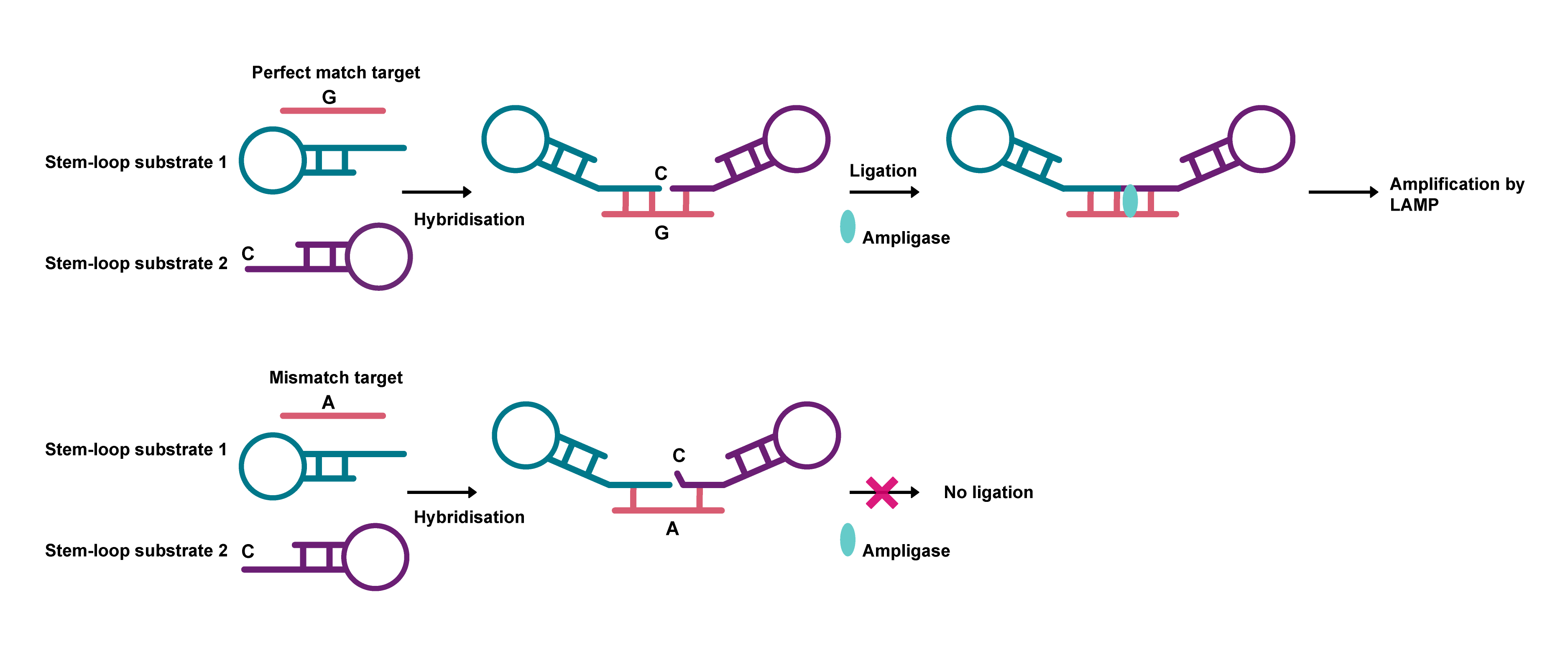
Figure 4. Representation of the LIEXA process. When there is a perfect match with the target sequence, the two loops are ligated, which enables a LAMP reaction.
LIEXA requires only a pair of universal primers and can be conducted under isothermal conditions, making it a potentially transformative tool for a wide array of biomedical research and diagnostic applications.
By simplifying the detection process and reducing reliance on expensive and complex equipment, LIEXA could pave the way for more effective and personalised medical interventions.
Conclusion
Improved detection of single point mutations can greatly expand the opportunities in advanced medicine, including precision medicine, infectious disease responses and genetic testing.
These ligase-based approaches offer alternatives to traditional PCR-based methods, improving specificity without losing sensitivity. The simplicity of the amplification methods, such as ligase chain reaction and rolling circle amplification, makes the detection process more accessible for point-of-care testing. Combined with cutting-edge technologies like CRISPR/Cas12a and Pyrococcus furiosus Argonaute (PfAgo), these approaches provide a powerful toolset for identifying genetic variations with high accuracy.
As a highly effective thermostable DNA ligase, Ampligase enables these techniques to deliver high-performance results and drive innovation in molecular diagnostics.

Related content
- Optimising automated high-throughput pathogen detection workflows
- The rise of LAMP for rapid point-of-need testing
References
- Li Y, Wang X, Wang M et al. (2023) Advances in ligase-based nucleic acid amplification technology for detecting gene mutations: a review. Mol Cell Biochem 478, 1621–1631 doi: 10.1007/s11010-022-04615-w
- Schalling M et al. (1993) Direct detection of novel expanded trinucleotide repeats in the human genome. Nature Genetics 4, 135. doi: 10.1038/ng0693-135
- Cao G, Chen X, Deng Y et al. (2021) Single-nucleotide variant of PIK3CA H1047R gene assay by CRISPR/Cas12a combined with rolling circle amplification. Analytica Chimica Acta, 1182, 338943 doi: 10.1016/j.aca.2021.338943
- Chen L, Eriksson A, Weström S et al. (2022) Ultra-sensitive monitoring of leukemia patients using superRCA mutation detection assays. Nat Commun 13, 4033. doi: 10.1038/s41467-022-31397-y
- Lih CJ et al. (2017) Analytical Validation of the Next-Generation Sequencing Assay for a Nationwide Signal-Finding Clinical Trial. J Mol Diagn. 19(2): 313–327. doi: 10.1016/j.jmoldx.2016.10.007
- Yohe S and Thyagarajan B (2017) Review of Clinical Next-Generation Sequencing. Arch Pathol Lab Med (2017) 141 (11): 1544–1557. doi: 10.5858/arpa.2016-0501-RA
- Wang L, He R, Lv B et al. (2021) Pyrococcus furiosus Argonaute coupled with modified ligase chain reaction for detection of SARS-CoV-2 and HPV. Talanta. 227:122154. doi: 10.1016/j.talanta.2021.122154
- He R, Wang L, Wang F et al. (2019) Pyrococcus furiosus argonaute-mediated nucleic acid detection. Chem Commun (Camb) 55(88):13219–13222. doi: 10.1039/c9cc07339f
- Liu Z, Yang L, Wei Q et al. (2020) A novel ligase chain reaction-based electrochemical biosensing strategy for highly sensitive point mutation detection from human whole blood. Talanta. 216, 120966 doi: 10.1016/j.talanta.2020.120966
- Wang H, Wang H, Sun Y et al. A general strategy for highly sensitive analysis of genetic biomarkers at single-base resolution with ligase-based isothermally exponential amplification. Talanta, 212, 120754, (2020) doi: 10.1016/j.talanta.2020.120754
