

Designing oligonucleotides for in vivo use often requires introducing several modifications to overcome limitations in stability, specificity and binding strength.
For the last three decades, researchers have been exploring an alternative approach that avoids these problems. Peptide nucleic acids (PNAs) are a unique class of molecules that combine the base-pairing capabilities of DNA with the robustness of peptides. These advantages could make PNAs indispensable in developing new classes of nucleic acid therapeutics and molecular diagnostics.
This post explores what PNAs are, why they are special, and how they are being applied in developing gene editing, new antibiotics and much more.
How are PNAs different to DNA?
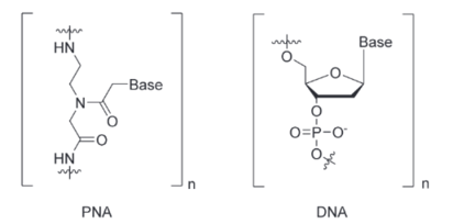
Figure 1. Chemical structure of PNA compared to DNA. PNA replaces the sugar–phosphate backbone of DNA with repeating N-(2-aminoethyl)-glycine units, which carries no charge.
PNAs are synthetic analogues of DNA in which the usual negatively charged sugar-phosphate backbone is replaced by a neutral, peptide-like backbone. This fundamental change provides PNAs with several useful properties:
- Strong and specific binding: Without the charge repulsion that exists between two DNA strands, a PNA can bind to a complementary DNA or RNA strand with higher affinity and specificity. Even a single base mismatch greatly destabilises a PNA–DNA duplex, which means PNAs excel at distinguishing single nucleotide differences. This allows developers to use shorter probes or antisense agents to achieve the same effect as longer DNA oligos.
- Strand invasion: PNAs can actively invade double-stranded DNA, displacing one strand to form a D-loop or forming a PNA2/DNA triplex via both Watson-Crick and Hoogsteen pairing.
- Enzymatic stability: The peptide-like backbone is not recognised by nucleases or proteases, so PNAs are highly resistant to enzymatic degradation. This allows them to circulate for longer in vivo without needing further modifications to increase nuclease resistance, unlike standard DNA or RNA therapeutics.
- Withstand harsh conditions: PNAs maintain their hybridisation stability across a wide range of temperatures and pH levels.1 This robustness makes them reliable in vivo and in assays that might involve extreme conditions.
- Neutral charge and no ion requirement: DNA/RNA binding often depends on salt concentrations to shield the negative charges. PNAs, being uncharged, work well even in low ionic strength environments and avoid issues with electrostatic interactions.1
While these benefits are valuable, PNAs also come with challenges. Their uncharged nature means PNAs don’t naturally penetrate cell membranes and have low solubility in aqueous solutions. However, there are several ways around this, such as attaching cell-penetrating peptide tags or using nanoparticle delivery systems.1
PNAs in therapeutic applications
PNAs were first invented in 1991 by Peter Nielsen and colleagues with clear applications in developing novel therapeutics.2
An antisense PNA can bind to a complementary mRNA (or pre-mRNA) and physically block its translation or alter its splicing, effectively silencing a gene. Unlike some other antisense oligonucleotides (ASOs), PNAs do not trigger RNase H cleavage of the RNA, instead working by steric hindrance and blocking cellular machinery.1
PNAs can also bind double-stranded DNA by strand invasion or triplex formation to prevent gene transcription. These mechanisms open the door to targeting virtually any gene or non-coding RNA involved in disease.
While no PNA-based drug has reached the market yet, there is significant research into developing these approaches and successfully delivering the molecules to their target in vivo.
Targeting genetic diseases and clinical testing
The high specificity of PNAs makes them attractive tools for tackling diseases with known genetic mutations or repeat expansions. The company NeuBase Therapeutics developed two PNA-based drug candidates that showed success in preclinical studies:
- NT-0100 knocked down mutant huntingtin protein levels in the brains of a Huntington’s disease mouse model.
- NT-0231.F was designed to treat myotonic dystrophy type 1 by binding to mutant DMPK mRNA to release trapped splicing factors. Preclinical tests in mouse models showed long-lasting distribution across different tissues and 70% reduction in muscle stiffness.
While NeuBase ceased operations due to financial constraints, these candidates demonstrated the potential for PNAs to hit targets that other approaches have struggled against.
For example, in August 2025, a PNA-based inhibitor of JAK2 received FDA Orphan Drug Designation for treating polycythaemia vera, a genetic disorder that causes an overproduction of blood cells.
Some PNA oligos have shown promising results in clinical trials, including EOM613, which is a candidate immune modulator aiming to prevent harmful cytokine storms. In a phase 1/2a trial of hospitalised patients with severe COVID-19, the PNA oligo led to lower pro-inflammatory cytokines and reduced inflammation.
EOM613 has also been tested in phase 2 trials of cachexia (severe muscle loss), showing improvements in patients with cancer or AIDS.3
Gene editing with PNAs
PNA oligos are also proving to be valuable tools in gene editing. Thanks to their strand invasion abilities, PNA oligos can trigger recombination and repair mechanisms that allow an ssDNA template to incorporate a genomic edit.
To maximise strand invasion, PNA designs, such as bis-PNA and tail clamp PNA, incorporate a flexible linker sequence to bind the target in parallel and antiparallel simultaneously, forming a triplex structure.4 This phenomenon works best when targeting a polypurine sequence near the modification of interest.
The editing relies solely on the cell’s natural repair processes unlike CRISPR, where the bacterial Cas9 enzyme needs to be added to create DNA strand breaks.
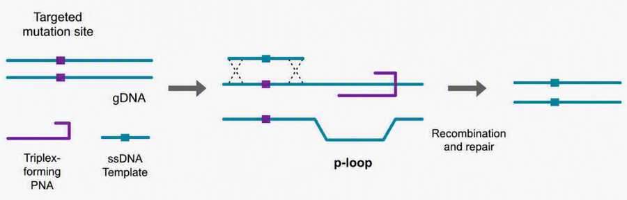 Figure 2. Representation of how tail-clamp PNA can invade the DNA double helix and initiate recombination and repair processes to introduce gene edits.
The PNA and ssDNA template can be co-delivered with nanoparticles, allowing administration in vivo. In 2025, researchers used PNA to modify mutations that cause cystic fibrosis in mice in utero.4 The gene editing persisted throughout the body and into adulthood.
This approach has also been investigated for treating other monogenic disorders, such as beta-thalassemia, sickle cell disease, Huntington’s disease and Duchenne muscular dystrophy.5
Fighting infections with antisense PNAs
One interesting therapeutic use of PNAs is for precision antibiotics. Pathogens, especially multidrug-resistant bacteria, can potentially be tackled by PNAs that target essential bacterial genes.
Even short PNAs of 9–12 bases can silence genes by binding to the mRNA and resulting in specific growth inhibition of the bacteria. This approach can, in theory, hit any bacterial gene, circumventing conventional drug resistance mechanisms.
In one example, researchers linked PNAs to cell-penetrating peptides (CPPs) to help them get inside the cells of carbapenem-resistant Acinetobacter baumannii, a notorious hospital superbug.1 The PNA was programmed to bind the start codon region of essential genes. These PNA–CPP conjugates killed the target bacteria without affecting unrelated bacterial species and, in cell culture, they showed no toxicity to human cells at effective doses.
In a recent study, a team from McGill University used antisense PNAs conjugated to CPPs to target essential genes in multidrug-resistant Salmonella.6 These 10-base long oligos were effective in vivo using C. elegans as an infection model, improving survival for the nematode worm.
These gene silencing antibiotics are still in the research stage, but they indicate how PNAs could form highly specific drugs to target a single strain of bacteria and overcome resistance.2
PNAs in cancer therapeutic development
Researchers have tested PNAs targeting oncogenes and oncomiRs (oncogenic microRNAs) in cancer models.
In 2025, researchers described the development of an antisense PNA targeting mRNA coding for PD-L1, a widely upregulated protein in cancers. The PNA oligo, synthesised using monomers from LGC Biosearch Technologies, was delivered into SK-OV3 cancer cells using an oncolytic adenovirus. This led to a significant downregulation of PD-L1, which is a key target for improving the effectiveness of cancer immunotherapies.7
In another study, a PNA designed against microRNA-21 (a microRNA that promotes cancer growth) was able to curb the growth and spread of breast cancer cells in vitro and even suppress tumour progression in mice.8
Additionally, PNAs can be directed to block transcription of specific genes, such as MYCN, to halt the growth of certain cancers.9 This works by being conjugated to a nuclear localisation signal and binding tightly to the DNA coding strand.
PNA-powered molecular diagnostics
The strong, specific binding that makes PNAs great for therapy is equally valuable in molecular diagnostics. PNAs are already a key component in several innovative diagnostic techniques, providing accuracy and speed that standard DNA probes struggle to match.
One PNA-based diagnostic tool is PNA-FISH (Fluorescence In Situ Hybridisation), where fluorescently labelled PNAs act as probes to detect microbial RNA or DNA in clinical samples. Because PNAs bind with high affinity and penetrate biological samples well, PNA-FISH can deliver quick, reliable results. For example, PNA-FISH assays have been designed to identify specific bloodstream infections.10
Beyond FISH, PNAs are also used in PCR clamping to improve mutation detection. This is where a PNA oligomer binds tightly to the wild-type DNA sequence, preventing DNA polymerase from amplifying it. If the DNA carries even a single-base mutation, the PNA does not bind, allowing the mutant sequence to be preferentially amplified.
PCR clamping with PNAs has been applied to identify low levels of clinically important mutations, such as those in the KRAS gene, which could help guide targeted cancer therapies.11,12
PNAs can even be used to detect biomarkers, such as microRNAs, more easily. For instance, researchers created PNA-based biosensors that fluoresce in the presence of a microRNA associated with prostate cancer.13
Unlike RT-PCR, these biosensors don’t require enzymatic amplification thanks to PNA’s strong binding affinity. The specificity, sensitivity and stability of PNAs help simplify protocols by removing the need for stringent conditions or thermal cycling. PNAs can even be used in blood or serum-based tests without nucleases degrading the probe.
As these PNA-based diagnostic tools continue to develop, they could enable faster and more precise results for point-of-care testing and personalised medicine.
Building PNA oligos
PNAs are synthesised in a manner very similar to how we produce peptides or DNA oligonucleotides. Typically, PNAs are made via automated solid-phase synthesis using the chemistry of peptide bond formation.
Each PNA monomer (which corresponds to a nucleobase) is added sequentially to a growing chain bound on a solid support. Instead of the phosphodiester linkage, an amide bond is formed between monomers, using coupling reagents much like those in peptide synthesis.
The bases on PNA monomers must be protected during synthesis since the usual DNA protecting groups won’t work in the peptide synthesis environment. After extensive screening, we have found that benzhydryloxycarbonyl (Bhoc) is the best choice for protecting the exocyclic amino groups of the nucleobases. The milder chemistry allows the synthesis of PNA with sensitive reporter groups, while an Fmoc group is used to protect the primary amino group.
In addition to the four Fmoc/Bhoc monomers, we offer a hydrophilic spacer molecule, AEEA, which increases the solubility of PNA, is used in bis-PNA and can act as a pseudo 5’ when labelling PNA with biotin, ROX, TAMRA, etc.
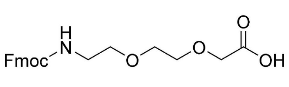
Figure 3. AEEA-OH Fmoc spacer. Useful for reducing the hydrophobicity of PNAs and introducing flexible linkers for bis-PNA and tail clamp PNAs.
The availability of high-quality PNA monomers means that labs can custom-make PNAs tailored to their target of interest.
For more details about PNA synthesis and labelling, read our education and support articles:
The value of PNAs
PNAs offer unique advantages where traditional oligos fall short and these are now being translated into real-world applications. PNA-based therapeutics are still largely in the experimental or preclinical stage, but the breadth of research shows their potential from antibiotics to cancer therapy.
PNAs exemplify how chemistry can expand biology’s toolkit, transitioning from a curious lab invention into a versatile platform for innovation in both therapy and diagnostics. With each new study demonstrating the value of PNAs, the case grows stronger that PNAs will play a key role in the future of medicine and biotechnology.
Take a look at our range of PNAs, among our 1,000 different nucleic acid products available.

References
- McLelland V et al. (2023) Therapeutic and diagnostic applications of antisense peptide nucleic acids. Mol Ther Nucleic Acids 35(1):102086 doi: 10.1016/j.omtn.2023.102086
- Nejad AJ et al. (2021) Targeting of the Essential acpP, ftsZ, and rne Genes in Carbapenem-Resistant Acinetobacter baumannii by Antisense PNA Precision Antibacterials. Biomedicines 9(4):429 doi: 10.3390/biomedicines9040429
- Nielsen PE, Egholm M, Berg RH, Buchardt O (1991) Sequence-selective recognition of DNA by strand displacement with a thymine-substituted polyamide. Science 254(5037):1497-500
- Chasen M, Hirschman SZ, Bhargava R (2011) Phase II study of the novel peptide-nucleic acid OHR118 in the management of cancer-related anorexia/cachexia. J Am Med Dir Assoc. 12(1):62-7. doi: 10.1016/j.jamda.2010.02.012
- Ricciardi AS et al. (2025) Systemic in utero gene editing as a treatment for cystic fibrosis. PNAS 122(24):e2418731122 doi: 10.1073/pnas.2418731122
- Eltaweel N et al. (2025) Gene editing using gamma modified PNA: HBB gene as a model. Journal of Genetic Engineering and Biotechnology 23(4):100562 doi: 10.1016/j.jgeb.2025.100562
- El Fateh M et al. (2025) Antibacterial activities of novel peptide nucleic acids targeting Salmonella penicillin-binding proteins. Mol Ther Nuc Acids 36(4):102762 doi: 10.1016/j.omtn.2025.102762
- Falanga AP et al. (2025) Engineering an oncolytic adenoviral platform for precise delivery of antisense peptide nucleic acid to modulate PD-L1 overexpression in cancer cells. International Journal of Pharmaceutics 668:124941 doi: 10.1016/j.ijpharm.2024.124941
- Yan LX et al. (2011) Knockdown of miR-21 in human breast cancer cell lines inhibits proliferation, in vitro migration and in vivo tumor growth. Breast Cancer Res 13, R2 doi: 10.1186/bcr2803
- Tonelli R et al. (2005) Anti-gene peptide nucleic acid specifically inhibits MYCN expression in human neuroblastoma cells leading to cell growth inhibition and apoptosis. Mol Cancer Ther 4 (5): 779–786 doi: 10.1158/1535-7163.MCT-04-0213
- Sungho Kim et al. (2024) Fast and accurate multi-bacterial identification using cleavable and FRET-based peptide nucleic acid probes. Biosensors and Bioelectronics doi: 10.1016/j.bios.2024.116950
- Oh JE et al. (2010) Detection of Low-Level KRAS Mutations Using PNA-Mediated Asymmetric PCR Clamping and Melting Curve Analysis with Unlabeled Probes. J Mol Diag 12(4):418-424 doi: 10.2353/jmoldx.2010.090146
- Shimane G et al. (2024) Molecular diagnosis for detecting KRAS mutation in peritoneal washing fluid of pancreatic ductal adenocarcinoma. Sci Rep 14, 21732 doi: 10.1038/s41598-024-72569-8
- Metcalf GAD et al. (2016) Amplification-Free Detection of Circulating microRNA Biomarkers from Body Fluids Based on Fluorogenic Oligonucleotide-Templated Reaction between Engineered Peptide Nucleic Acid Probes: Application to Prostate Cancer Diagnosis. Anal. Chem. 88(16):8091–8098 doi: 10.1021/acs.analchem.6b01594
