
Updated : Tue, January 25, 2022 @ 10:23 PM
This is the first blog post in a two-part series highlighting how Black Hole Quencher™ dyes can be incorporated into assays to enable novel and highly sensitive detection and imaging applications.
Quenchers absorb excitation energy from a fluorophore, such as a dye, suppressing their emission of fluorescent signal when in close proximity. When the fluorophore and quencher are separated, the quencher can no longer absorb the emission from the fluorophore, enabling fluorescence to be detected. Black Hole Quencher (BHQ™) dyes are the original true dark quenchers. They have no fluorescent signal themselves and efficiently deliver results through a combination of FRET and static quenching, avoiding the residual background signal common to other quenchers, such as TAMRA.
|
The history of LGC, Biosearch Technologies and PCR stretches back to the 1980s, when Kary Mullis – under the encouragement of Ron Cook (founder of Biosearch) – used a Biosearch SAM I DNA synthesizer to create oligos for use in his experiments, eventually resulting in the discovery of PCR. Since the introduction of BHQ dye technology in 2000, they have served as the quencher of choice for qPCR probes and other fluorescence-quenched probe applications. The use of Dual-labeled BHQ Probes in quantitative real-time PCR |
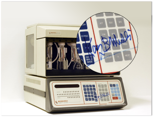 In the 1980s, Biosearch developed and manufactured automated, |
Quantitative real-time PCR (qPCR) offers affordable, specific and high-throughput measurement of nucleic acids, with the option to multiplex. There are two approaches for real-time visualisation of amplified DNA products – non-specific fluorescent DNA dyes and fluorescently labelled oligonucleotide probes. Fluorescent probes have proved to be the superior option due to higher specificity and lower susceptibility to detecting primer dimers1, resulting in BHQ dyes becoming almost ubiquitous in qPCR applications. Six out of seven of the World Health Organization’s COVID-19 testing protocols use BHQ dyes and the top ten molecular diagnostic manufacturers leverage BHQ technology in their assays.
BHQ dyes are easy to use and simplify the design, implementation, and interpretation of qPCR assays. They are integral to pathogen detection as well as assays that query single and multiplex gene expression, copy number variation, SNP genotyping and presence/absence. BHQ dyes have revolutionised the ability to multiplex and have demonstrated themselves to be invaluable in a pandemic, such as the 2009 H1N1 Flu pandemic and the current SARS-CoV-2 pandemic.
Dual-labeled BHQ Probes are widely used in end-point PCR too. Here are two examples of how they’ve been used during the SARS-CoV-2 pandemic:
BHQ Probes enable ultra-high-throughput testing for COVID-19
To meet the demand for large-scale COVID-19 testing, the Biosearch Technologies ultra-high-throughput workflow (including the NexarTM industrial scale, automated end-point PCR testing platform) was combined with SARS-CoV-2 PCR primers and BHQ Probes. This was implemented within some of the UK’s laboratory network, with each system enabling up to 150,000 samples a day to be tested – the highest PCR testing capacity per day per instrument system worldwide. The BHQ Probes are an essential component of the system, with fully automated fluorescence detection on the ArayaTM.
BHQ dyes and digital PCR used for wastewater surveillance of SARS-CoV-2 in the community
Digital PCR (dPCR) has seen a considerable technical progression over the last decade, with a vast range of new applications. dPCR uses the same core reagents as qPCR, but the end-point fluorescence is measured to detect and quantify the presence of a target sequence in each partition. The digital MIQE guidelines update in 2020 recommend that where hydrolysis probes are used, these should ideally be nonfluorescent quenchers2, such as BHQ dyes.

Innovative applications for BHQ dyes
Black Hole Quencher dyes aren’t just for use in dual-labelled probes. See below for examples of BHQ dyes making a strong contribution to cutting edge research:
BHQ dyes enable ultra-fast cycling for multiplexed cellular fluorescence imaging. Analysis of cancer cells using molecular techniques is essential for patient diagnoses and treatment plans, however current staining protocols require hours to days of sample processing. A method was designed using BHQ dyes and click chemistry for rapid repeated staining cycles of tumour cells and their surrounding environments sampled using fine needle aspiration. A fluorescent dye is bound to antibodies specific for particular markers on the tumour cells. If the target marker is present, the antibody will bind and fluorescence is detected. Instead of using harsh chemicals to remove or bleach the dye before restaining with a dye for another marker, the fluorescence is switched off using a Black Hole Quencher dye. The quencher has a tetrazine (Tz) attached and the fluorophore has a trans-cyclooctene (TCO) linking it to the antibody. When the quencher is added to the sample the Tz and TCO click together, immediately bringing the BHQ dye and the reporter dye in close proximity, thereby rapidly quenching the fluorescence. The sample is then ready to test for the next marker by adding another fluorescent antibody. The researchers were able to stain 12 markers within one hour, demonstrating that this method is highly efficient and gentle enough for multiplex profiling within a single sample.4 The addition of BHQ dye and click chemistry to this cycling method for cellular imaging demonstrates new possibilities for better cancer patient diagnostics and assessment of treatments.
Quantum dot nanobeacons with conjugated BHQ dye for single RNA labelling and imaging in live cells. Methods for detecting and imaging RNAs in live cells are in high demand, but techniques such as real-time quantitative PCR and Northern blotting require RNA extraction steps, so are not suitable for in vivo detection. In a recent study, a CdTe: Zn2+ quantum dot conjugated to BHQ1 and phosphorothioate co-modified DNA was shown to be capable of labelling and imaging single RNA in live cells. Detection occurs when the stem-loop hairpin DNA hybridises to target nucleic acid sequences, allowing the FRET-based nanobeacon to fluoresce.
To demonstrate live imaging in HIV-1 integrated cells, quantum dot nanobeacons (QD-NBs) were incubated with TNF-α activated U1 cells for 48 hours, then observed using a spinning disk confocal microscope. Live imaging of low-abundance nucleic acids in live cells was achieved by repeating this method but using shorter activation times, with 6 hours still resulting in fluorescent signals, indicating excellent sensitivity. To investigate an additional application of the QD-NBs, they were hybridised to HIV-1 genomic RNAs and packed into virions. This enabled real-time tracking of the viral uncoating process, viewed with spinning disk confocal microscopy. The results indicate that the QD-NB with conjugated BHQ dye is a highly sensitive new platform for tracking RNAs in live cells.5
An oligo and BHQ screening platform uncovered a small molecule inhibitor for ribonuclease P, contributing to the battle to find new antibiotics. RNA-protein complexes and RNA-processing enzymes are essential for microbial metabolism, making them appealing as antibiotic targets. Researchers developed a novel, real-time fluorescence-based assay with BHQ dye to investigate inhibitors for ribonuclease P (RNase P), a ribonucleoprotein ribozyme involved in 5’ tRNA processing. Two substrates for RNase P were designed utilising a FRET mechanism – the first a 24-nucleotide minihelix and the second a 77-nucleotide bipartite pre-tRNA, both coupled to a BHQ dye and fluorophore. For initial screening, 2560 compounds dissolved in DMSO were incubated with the minihelix substrate and active RNase P. Fluorescence was measured every 15 seconds for at least 5 minutes and Michaelis-Menten kinetics observed. Four potential positive compounds were then tested for inhibitory activity using the bipartite pre-tRNA substrate, in silico analysis and x-ray crystallography, revealing one compound – purpurin – to be the most promising inhibitor. Experimental data showed the minihelix assay to be reproducible, consistent with existing data on known RNase P inhibitors, tolerant of DMSO and suitable for high-throughput screening. The bipartite pre-tRNA substrate was used as a pre-tRNA analog and was demonstrated to have a comparable affinity. This screening assay utilising BHQ dye offers a novel method to find new compounds capable of inhibiting RNase P, potentially expanding the number of leads for antibiotics for clinical use.6
Check out the second blog post in this series where we continue exploring additional applications of BHQ dyes, such as use in microfluidic chips, aptamer probes and dual-labelled probes with locked nucleic acids.
References
- Kralik P and Ricchi M. A Basic Guide to Real Time PCR in Microbial Diagnostics: Definitions, Parameters, and Everything. Frontiers in Microbiology, Volume 8. https://doi.org/10.3389/fmicb.2017.00108. Published 2017. Accessed November 11, 2021.
- The dMIQE Group and Huggett JF. The Digital MIQE Guidelines Update: Minimum Information for Publication of Quantitative Digital PCR Experiments for 2020. Clinical Chemistry, Volume 66, Issue 8, 1012-1029. https://doi.org/10.1093/clinchem/hvaa125. Published 2020. Accessed November 11, 2021.
- Ciesielski M, Blackwood D, Clerkin T, Gonzalez R, Thompson H, Larson A, Noble R. Assessing sensitivity and reproducibility of RT-ddPCR and RT-qPCR for the quantification of SARS-CoV-2 in wastewater. Journal of Virological Methods, Volume 297. https://doi.org/10.1016/j.jviromet.2021.114230. Published 2021. Accessed November 5, 2021.
- Ko J, Oh J, Ahmed MS, Carlson JCT, Weissleder R. Ultra-fast Cycling for Multiplexed Cellular Fluorescence Imaging. Angewandte Chemie, Volume 132, Issue 17, 6697-7002. https://doi.org/10.1002/ange.201915153. Published 2020. Accessed November 1, 2021.
- Ma Y, Mao G, Huang W, Wu G, Yin W, Ji X, Deng Z, Cai Z, Zhang XE, He Z, Cui Z. Quantum Dot Nanobeacons for Single RNA Labeling and Imaging. Journal of the American Chemical Society, Volume 141, Issue 34, 13454-13458. https://doi.org/10.1021/jacs.9b04659. Published 2019. Accessed November 5, 2021.
- Madrigal-Carrillo EA, Díaz-Tufinio CA, Santamaría-Suárez HA, Arciniega M, Torres-Larios A. A screening platform to monitor RNA processing and protein-RNA interactions in ribonuclease P uncovers a small molecule inhibitor. Nucleic Acids Research, Volume 47, Issue 12, 6425-6438. https://doi.org/10.1093/nar/gkz285. Published 2019. Accessed November 8, 2021.